
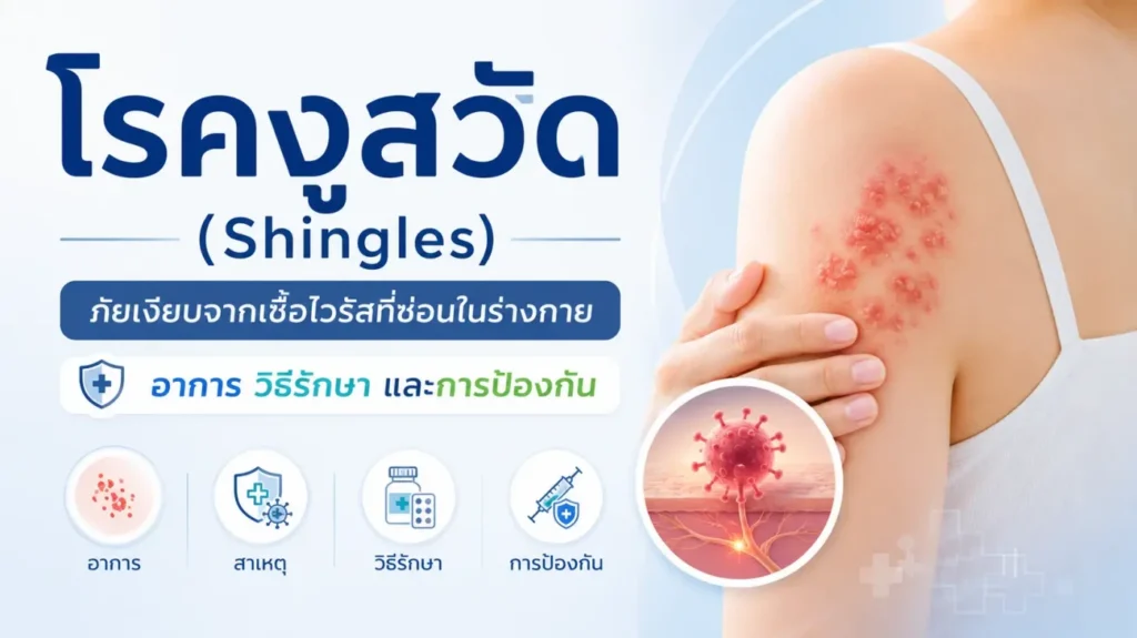
Most people mistakenly believe that “herpes zoster” (shingles) is merely a skin disorder with a rash or blisters that erupt for a few days and then resolve once crusted over. In reality, many patients continue to suffer from severe, persistent burning pain for months or even years, long after the external skin lesions have completely healed.
This type of neuropathic pain is known as Postherpetic Neuralgia (PHN), which is recognized as one of the most treatment-refractory and distressing forms of nerve pain (Neuropathic pain). It poses critical distress to patients, particularly elderly populations, where the pain frequently disrupts sleep cycles, interferes with activities of daily living, and induces long-term negative psychological sequelae. Today, the field of pain medicine has shifted from the traditional view of PHN as a mere dermatologic condition toward a shared understanding that it is a disease directly involving “damage and maladaptive neuroplasticity of the nervous system,” spanning the peripheral nerves, spinal cord, and higher brain centers.
Q&A: Etiopathogenesis, Prodromal Pain, and Mechanisms of Neural Destruction
What pathogen causes shingles, and where does it lie dormant in the body?
Herpes zoster (Shingles) is caused by the reactivation of the Varicella-Zoster Virus (VZV), the same agent responsible for primary chickenpox during childhood. Following clinical resolution of chickenpox, the virus is not entirely eradicated from the host. Instead, it migrates to establish a lifelong, latent infection within the Dorsal Root Ganglion (DRG)—a primary processing station for sensory neurons—as well as major cranial nerve ganglia, such as the ophthalmic and sensory branches of the Trigeminal nerve.
When the host experiences advancing age, waning cell-mediated immunity, sleep deprivation, cumulative stress, or pharmacologic immunosuppression, the dormant virus reactivates and rapidly replicates. It travels anterograde along sensory nerves to the skin, triggering characteristic vesicular eruptions and severe, localized pain. Consequently, lesions typically manifest unilaterally across a discrete cutaneous nerve distribution (Dermatome).
What are the clinical characteristics of “prodromal pain,” and why is it frequently misdiagnosed?
In many patients, active viral-mediated neural inflammation generates severe neurogenic pain approximately 2 to 7 days before the initial cutaneous vesicular eruption appears. Known as the prodromal phase, this phenomenon frequently leads to diagnostic confusion and delayed recognition, as it is often mistaken for:
- Acute myofascial back strain or lumbago
- Degenerative joint disease, arthralgia, or acute shoulder capsulitis
- Intercostal pleurisy or rib pain (frequently confounded with acute cholecystitis or biliary colic)
- Odontogenic pain or pulpitis (when reactivation occurs along the V2/V3 branches of the Trigeminal nerve)
- Acute coronary syndrome presenting with retrosternal chest pain radiating to the upper extremity
Why does herpes zoster generate such excruciating, intractable pain?
The pain does not stem solely from cutaneous blistering, but rather from direct structural destruction of the nervous system induced by active VZV replication. The pathology entails acute ganglioneuritis, marked ganglion swelling, extensive breakdown of the insulating protective sheath (Myelin sheath), and aberrant electrical hyperexcitability with ephaptic crosstalk.
When this inflammatory cascade remains sustained, it drives neuroplastic maladaptation within the dorsal horn of the spinal cord and supra-spinal nuclei into a state of Central Sensitization—effectively encoding a “centralized neural memory of deep-seated pain.” Patients subsequently endure deep, continuous burning discomfort, lancinating paroxysmal shock-like pain, and prominent Allodynia (severe mechanical hypersensitivity). In this state, innocuous, sub-threshold stimuli (such as the glide of lightweight clothing, a gentle breeze, or light water flow during showering) trigger massive aberrant discharges, provoking excruciating pain that often leaves patients unable to tolerate routine clothing or underwear.
Clinical Definition of Postherpetic Neuralgia (PHN) and Key Risk Criteria
In clinical practice, a diagnosis of Postherpetic Neuralgia (PHN) is established based on the following benchmark:
⏱️ Clinical Diagnostic Criteria
Persistent neuropathic pain enduring for more than 3 months following the initial onset of the acute dermatomal herpes zoster rash.
This persistent burning pain and mechanical allodynia can persist for months, years, or become a lifelong chronic neuropathic condition that severely impairs overall health-related quality of life. High-risk patient populations predisposed to transitioning from acute zoster to chronic PHN include:
- Advanced chronological age (impaired intrinsic neuro-regenerative capacity)
- High baseline visual analog/numeric pain scores during the first week of onset
- Extensive cutaneous vesicular eruption with confluence or epidermal necrosis
- Prominent early presence of cutaneous allodynia
- Underlying comorbidities, such as poorly controlled diabetes mellitus, systemic immunocompromise, chronic distress, and prolonged sleep deprivation
Emergency Management Principles: The Critical 72-Hour “Golden Window”
The primary clinical priority in managing acute herpes zoster is the immediate initiation of oral antiviral therapy within the 72-hour golden window of initial rash emergence. Timely administration of standard antiviral agents (such as Acyclovir, Valacyclovir, or Famciclovir) during this critical period provides key therapeutic benefits:
- Halts viral replication and limits neurotropic dissemination across the neuraxis
- Suppresses the acute neuroinflammatory response and attenuates neural edema
- Preserves structural integrity of the Dorsal Root Ganglion (DRG) from irreversible injury
- Reduces the severity, formation of new vesicles, and duration of cutaneous lesions
- Significantly reduces the incidence and transition rate toward chronic Postherpetic Neuralgia (PHN)
Modern pain medicine has evolved past the historical doctrine of “observing skin lesion healing before addressing chronic pain” to an aggressive paradigm of “rapid, assertive suppression of acute neuropathic pain within the first 72 hours” to arrest central maladaptive remodeling and preempt permanent Chronic Neuropathic Pain.
Neuromodulatory Pharmacotherapy Protocols
When patients transition into established PHN, standard systemic analgesics are generally ineffective. Specialist pain teams employ multimodal regimens targeted at restoring neurochemical equilibrium:
- Gabapentinoids (Voltage-gated calcium channel alpha-2-delta ligands): Including Pregabalin, Gabapentin, and Mirogabalin. These agents selectively modulate overactive voltage-gated calcium channels, attenuating downstream excitatory neurotransmitter release. Among these, the next-generation ligand Mirogabalin has garnered widespread clinical utility due to its distinct binding kinetics and high receptor selectivity. It provides robust neuropathic pain relief in PHN with a demonstrably lower incidence of treatment-limiting side effects, such as deep somnolence, cognitive fogging, and peripheral ataxia, particularly in geriatric populations.
- Central Descending Pain Pathway Modulators (Antidepressants): Including Amitriptyline, Nortriptyline, and Duloxetine. These agents augment endogenous descending inhibitory serotonergic and noradrenergic pathways from the brainstem to the dorsal horn of the spinal cord, while mild sedative actions serve to normalize broken sleep architecture.
- Topical Local Anesthetics: While Lidocaine Patch 5% was historically a first-line intervention for localized allodynia, regulatory and supply constraints have led to discontinuation of importation and sales across Thailand. Consequently, interventional pain physicians frequently utilize compounded eutectic mixtures of local anesthetics (EMLA Cream) applied as a thin layer over intact hyperalgesic dermatomes to induce reversible blockade of cutaneous nociceptive A-delta and C fibers. This significantly blunts burning pain and friction discomfort from clothing, enabling functional mobility, bathing, and routine activities of daily living.
- Critical Safety Warning: EMLA cream is strictly contraindicated on unhealed blisters, active open ulcers, or broken skin barriers. Furthermore, application over excessively large surface areas is prohibited to prevent systemic absorption, toxic plasma concentrations, and secondary local anesthetic systemic toxicity (LAST), including cardiotoxicity.
Minimally Invasive Interventional Pain Management
In cases refractory to oral pharmacotherapy where debilitating pain disrupts basic biological function, or where systemic pharmacotherapy causes intolerable adverse events (such as heavy sedation, ataxia, or fall risks), targeted interventional procedures are deployed:
- Intercostal Nerve Block: Targeted deposition of local anesthetic and adjuvant along affected intercostal nerves for dermatomal thoracic or abdominal wall zoster pain.
- Cervical / Lumbar Epidural Steroid Injection: Targeted delivery of non-particulate/particulate corticosteroids into the epidural space, bathing inflamed exiting nerve roots and hyperemic DRG structures to rapidly mitigate acute neuroinflammation.
- Stellate Ganglion Block (SGB): Sympathetic blockade targeting the cervical sympathetic chain for cephalic, cervical, or upper-extremity distributions to downregulate sympathetically maintained pain and augment microvascular perfusion.
- Dorsal Root Ganglion Pulsed Radiofrequency (DRG-PRF): Fluoroscopic- or CT-guided placement of radiofrequency cannulae directed precisely at the pathologic DRG, delivering non-neurodestructive bursts of high-voltage radiofrequency energy to establish an electric field that modulates hyperactive primary afferents, yielding durable, safe analgesia in chronic PHN.
- Spinal Cord Stimulation (SCS): For refractory, long-standing multi-year PHN failing conservative and intermediate interventions, neuromodulatory dorsal column electrode implantation delivers electrical impulses that disrupt aberrant ascending pain signaling networks, restoring functional independence.
🚨 Critical Clinical Red Flag: Herpes Zoster Ophthalmicus (HZO)
When vesicular eruptions appear along the forehead, peri-orbital rim, upper eyelid, or the tip/side of the nose (Hutchinson’s sign), accompanied by conjunctival injection, ocular pain, or decreased visual acuity, this constitutes an emergency within ophthalmology. This distribution indicates viral replication along the ophthalmic division (V1) of the Trigeminal nerve, carrying significant risk for severe neurotrophic keratitis, corneal stromal ulceration, corneal perforation, secondary acute glaucoma, and irreversible permanent visual loss or blindness. Immediate emergency ophthalmologic consultation, along with targeted topical and aggressive systemic antiviral therapy, is mandated.
Preventive Immunization Strategies with Modern Zoster Vaccines
In modern preventive medicine, immunoprophylaxis remains the definitive, most efficacious intervention to suppress herpes zoster reactivation and avert downstream PHN development:
Clinical Insights on the Shingrix® Vaccine
The contemporary gold-standard vaccine recommended internationally is Shingrix®, a Recombinant Zoster Vaccine (RZV) possessing key structural innovations superior to previous live-attenuated formulations:
- Non-Live Antigen Platform (Non-live vaccine): Composed of a recombinant viral surface glycoprotein E (gE) combined with the proprietary AS01B adjuvant system. This formulation exhibits an exceptional safety profile, allowing safe administration in immunocompromised hosts, oncology patients, and older adults in whom live-attenuated vaccines are contraindicated.
- Robust Clinical Efficacy: Rigorous large-scale clinical trials demonstrate sustained protective efficacy exceeding 90% against both herpes zoster reactivation and the subsequent onset of PHN across long-term follow-up intervals.
- Dosing Protocol: Administered as a two-dose intramuscular series, with the second dose scheduled 2 to 6 months after the primary dose.
Q&A: Frequently Asked Clinical Questions Regarding Vaccination
- Can a patient receive the Shingrix® vaccine immediately following clinical recovery from an acute shingles episode? For immunocompetent individuals, international consensus guidelines generally recommend waiting approximately 6 to 12 months after the acute rash has completely healed before initiating vaccination. An acute episode boosts endogenous cell-mediated immunity naturally, resulting in a low incidence of recurrence during the immediate first year. However, in select patient cohorts—such as advanced age, active malignancy, or established immunosuppression—clinicians may recommend vaccination earlier, once the active acute lesions have completely crusted and re-epithelialized, due to an elevated risk of recurrent viral reactivation.
- Is zoster vaccination still indicated if the individual received the childhood varicella (chickenpox) vaccine? Yes, it remains strongly indicated. The childhood varicella vaccine utilizes a live-attenuated viral strain (Oka strain). While highly protective against wild-type chickenpox, this attenuated virus retains the capacity to establish latent infection within dorsal root ganglia. Consequently, vaccine recipients remain susceptible to reactivation as zoster later in life during periods of immunosuppression (albeit with lower reported rates and reduced PHN severity relative to wild-type chickenpox infection). Administration of Shingrix in adults (>50 years of age) or high-risk populations provides definitive, target-specific humoral and cell-mediated immunological protection.
- What is the expected frequency and profile of post-vaccination adverse events? Because Shingrix® utilizes an active adjuvant (AS01B) to induce robust immune responses, reactogenicity is relatively common, though self-limiting and typically resolving within 2 to 3 days. Common transient adverse effects include: injection-site erythema, pain, local swelling, low-grade pyrexia, myalgia, fatigue, systemic malaise, chills, or headache. These reactogenic symptoms can be effectively managed with symptomatic antipyretics and analgesics, such as paracetamol.
References
- Liu Q, Han J, Zhang X. Peripheral and central pathogenesis of postherpetic neuralgia. Skin Research and Technology. 2024;30(8).
- Ding S, Wen S, Kang H, et al. Association of the incidence of postherpetic neuralgia with early treatment intervention of herpes zoster and patient baseline characteristics: A systematic review and meta-analysis of cohort studies.
- Aggarwal A, Suresh V, Gupta B, Sonthalia S. Post-herpetic Neuralgia: A Systematic Review of Current Interventional Pain Management Strategies.
- Singh S, Gupta R, Kaur S. Post-herpetic neuralgia: A review of current management strategies. Indian Journal of Pain. 2013;27(1):12.
- Sun CL, Li XL, Li CW, et al. High-voltage, Long-duration Pulsed Radiofrequency to the Dorsal Root Ganglion Provides Improved Pain Relief for Herpes Zoster Neuralgia in the Subacute Stage. Pain Physician. 2023.
- Australian Immunisation Handbook. Zoster (herpes zoster).
- Medscape Neurology. Postherpetic Neuralgia. Updated 2025.
- Herpes Zoster and Post-Herpetic Neuralgia- Diagnosis, Treatment, and Vaccination Strategies. 2024.
- CDC Shingles Vaccination Guidelines. Recombinant Zoster Vaccine (Shingrix).