Contemporary Facet-Mediated Pain Management: From Diagnosis to Precision Medicine
Although most patients are familiar with the condition known as “herniated disc,” in reality, the posterior spinal joint (Facet Joint or Zygapophyseal Joint)—a small joint connecting each adjacent vertebra—is an exceptionally common pain generator encountered in routine clinical practice.
Because the Facet Joint is a true synovial joint possessing a synovium, synovial fluid, and abundant sensory nerve endings, it is prone to inflammation, degeneration, and abnormal biomechanical loading just like the knee or hip joint. Epidemiological data indicate that pathology of this joint accounts for up to 15–45% of chronic low back pain and 40–60% of chronic neck pain in selected patient populations.
Clinical Challenge: The diagnosis of facet joint pain cannot rely solely on neuroimaging modalities such as MRI or CT scans. Many patients exhibit age-related degenerative changes on imaging that do not necessarily represent the active trigger of clinical pain. Consequently, diagnostic interventional procedures play a crucial role in accurately pinpointing the pain source.
Q&A: Facet Joint Injection and Medial Branch Block (MBB)
1. What is a Facet Joint Injection, and who is an ideal candidate?
It is the direct intra-articular administration of a combination of a local anesthetic and a corticosteroid into the joint cavity under fluoroscopy or CT guidance. Currently, this targeted procedure yields superior outcomes in the following patient cohorts:
- Patients with acute or recent-onset pain.
- Patients with a strong clinical suspicion of acute facet synovitis/arthritis.
- MRI evidence demonstrating salient active lesions, such as peri-facet edema or increased STIR signal.
- Patients with contraindications or technical limitations to Radiofrequency Ablation (RFA).
2. What is a Medial Branch Block (MBB)?
The facet joint receives and transmits nociceptive signals via small nerve branches termed the medial branch nerves. Accordingly, a Medial Branch Block (MBB) involves the targeted injection of a local anesthetic onto these nerves to confirm the pain source. Presently, MBB is established as the gold standard for diagnosing facet-mediated pain and serves as a precise prognostic screening tool prior to proceeding with RFA.
Q&A: Paradigm Shift: From Diagnostic Block to Therapeutic MBB
Historically, medial branch blocks were classified purely as diagnostic procedures; significant pain reduction following the block served strictly as a prognostic indicator of favorable response to thermal radiofrequency neurotomy (RFA).
Today, this paradigm has evolved significantly. Clinical evidence and the latest Medicare Local Coverage Determinations (LCD) in the United States recognize Therapeutic Medial Branch Blocks in select patient subpopulations, particularly:
- Elderly patients or individuals with complex multiple comorbidities.
- Patients who are unsuitable for or have contraindications to RFA.
- Patients who previously demonstrated robust, durable relief from MBB and maintain functional pain control through scheduled periodic blocks.
Coverage Criteria (Medicare Guidelines): Therapeutic MBB is reimbursed as an ongoing management strategy if the index block achieved at least a 50% reduction in pain or functional improvement with analgesia persisting for 3 months or longer, limited to a maximum of 4 sessions per spinal region per rolling 12-month period.
Q&A: Clinical Technique and Positive Block Diagnostic Criteria
Should a Diagnostic Block be performed once or twice (Single vs. Dual Block)?
This remains a point of active discussion across international pain societies:
- Spine Intervention Society (SIS) & ASIPP: Recommend comparative dual blocks to minimize the rate of false-positive outcomes.
- Contemporary Consensus Guidelines: Acknowledge that a single diagnostic block is clinically pragmatic and sufficient if the index block yields an unequivocal, profound response of >75–80% pain relief. A single-block protocol limits procedural morbidity, lowers healthcare expenditures, and accelerates transition to definitive therapeutic management.
Fluoroscopic vs. Ultrasound Guidance: Comparative Utility
- Fluoroscopy (Gold Standard): Remains the primary modality for lumbar facet interventions due to clear visualization of osseous architecture and anatomic landmarks, superior needle placement precision, extensive validation across clinical literature, and immediate seamless workflow transition to RFA.
- Ultrasound-Guided: Offers zero ionizing radiation exposure, real-time visualization of adjacent soft tissue and vascular anatomy, and lower operational cost. Although non-inferior clinical outcomes are reported in literature, the technique remains heavily operator-dependent and technically challenging in patients with high body mass index (BMI) or severe spinal deformities.
Advanced Interventions and Emerging Therapies
Radiofrequency Ablation (RFA)
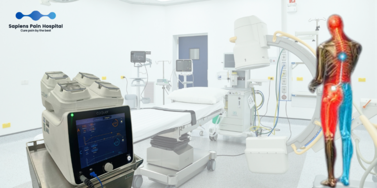
Following a validated positive diagnostic MBB, patients are indicated for thermal radiofrequency neurotomy. This minimally invasive intervention denatures the nociceptive pathways of the medial branch nerves without disrupting native structural joint biomechanics or requiring open surgery, conferring durable analgesia lasting an average of 8–12 months.
Regenerative Medicine (PRP & Stem Cells)
Because the facet joint is a true diarthrodial synovial joint, orthobiologic regenerative strategies have garnered substantial clinical interest over recent years:
- Platelet-Rich Plasma (PRP): Recent systematic reviews identify moderate-level evidence supporting PRP for axial facetogenic pain. Several clinical trials indicate sustained long-term symptom relief superior to intra-articular corticosteroids; however, it remains an evolving modality and does not currently supplant established interventional paradigms like MBB or RFA.
- Mesenchymal Stem Cells (MSCs): Clinical evidence remains sparse and strictly exploratory, warranting robust, high-quality randomized controlled trials prior to routine integration.
References
- Cohen SP, Bhaskar A, Bhatia A, et al. Consensus practice guidelines on interventions for lumbar facet joint pain from a multispecialty, international working group. Reg Anesth Pain Med. 2020;45:424-467.
- Manchikanti L, Kaye AD, Soin A, et al. A Systematic Review of Regenerative Medicine Therapies for Axial Spine Pain of Facet Joint Origin. Curr Pain Headache Rep. 2025.
- Nisolle ML, Ghoundiwal D, Engelman E, et al. Comparison of ultrasound-guided versus fluoroscopy-guided medial lumbar branch block. BMC Anesthesiol. 2023;23:76.
- Levin J, Barrette K, Ghaffari C, et al. Intra-articular cervical facet joint corticosteroid injections in patients with increased peri-facet MRI STIR signal. Interv Pain Med. 2025;4:100646.
- Geoffroy M, Beissat M, Kanagaratnam L, et al. Platelet-rich plasma versus corticosteroids in facet joint syndrome: A controlled, randomized, double-blind study. Joint Bone Spine. 2026;93:106001.
- Baltzer AW, Enneper J, Baltzer LM, Godde G. Platelet Rich Plasma for the Therapy of the Lumbar Facet Joint Syndrome. Orthop Rev (Pavia). 2025;17:141416.
- Medicare Local Coverage Determination (LCD): Facet Joint Interventions for Pain Management (L38773), revised July 2024.
- Manchikanti L, Pampati V, Cash KA, et al. Lumbar facet joint nerve blocks in managing chronic facet joint pain. Pain Physician. 2008;11:121-132.