
Historically, when discussing chronic neck or back pain, the primary focus has predominantly centered on herniated intervertebral discs compressing spinal nerves. However, another crucial, highly prevalent, and frequently overlooked etiology is “Facet Joint Pain”.
Currently, an innovative intervention known as Radiofrequency Ablation (RFA) of nerves has emerged as a vital therapeutic bridge, filling a crucial gap for patients whose “symptoms are not severe enough to warrant major spinal surgery, yet do not sufficiently respond to conservative management.”
Q&A: Facet Joint Anatomy and Pain Pathomechanics
What is a Facet Joint?
Facet joints are paired, small synovial joints located bilaterally on the posterior aspect of each vertebral motion segment. They stabilize and guide physiological ranges of spinal motion—such as flexion, extension, lateral bending, and axial rotation—maintaining spinal biomechanical balance. These joints span along the entire spinal column, from the cervical and thoracic regions down to the lumbar spine. Structurally composed of articular cartilage, a fibrous capsule, and abundant nociceptive nerve endings, inflammation or degenerative arthropathy here can elicit chronic, disabling pain comparable to advanced osteoarthritis of the knee.
Why is the Incidence of Facet Joint Arthropathy Rising in the Modern Population?
A primary accelerating factor is prolonged forward neck flexion and slouched posture during occupational activities, commonly referred to as Text Neck Syndrome. The adult human head typically weighs approximately 4–6 kilograms; however, with progressive forward flexion angles, the biomechanical load and cumulative shear stress exerted on the intervertebral discs and posterior facet joints multiply exponentially. This mechanical overload precipitates accelerated joint degeneration, capsular hypertrophy/fibrosis, and chronic synovitis. This presentation is exceptionally common among clinicians performing sustained static forward-head procedures—such as dentists and surgeons—as well as desk workers, software engineers, graphic designers, and patients presenting with post-motor vehicle collision Whiplash Injury.
In-Depth Pathophysiological Mechanism
As intervertebral discs degenerate and lose disc height, compressive and torsional loading forces are redistributed posteriorly onto the facet joints. This leads to mechanical friction, capsular inflammation, joint effusion, and osteophyte formation. The affected joints transmit constant nociceptive signals via dedicated tiny nerve fibers termed the Medial Branch Nerves directly into the central nervous system. Protracted peripheral nociceptive barrage can induce neuroplastic alterations within the spinal cord and brain, ultimately culminating in Central Sensitization.
Level-Specific Clinical Pain Syndromes
- Cervical Facet Pain: Chronic axial neck pain radiating down into the trapezius and periscapular region, distinctly exacerbated by cervical extension and axial rotation. Pain often refers cephalad toward the occiput, retroauricular area, temple, or retro-orbital region, clinically mimicking Cervicogenic Headache or migraine, in the absence of true radicular dermatomal numbness or motor deficits.
- Thoracic Facet Pain: Deep, paravertebral thoracic pain between the scapulae, aggravated by trunk rotation, spinal flexion, or axial hyperextension, frequently accompanied by protective deep paraspinal muscular hypertonicity and spasm.
- Lumbar Facet Pain: Axial lower back pain referring into the buttocks, trochanteric region, or groin, with marked morning stiffness or pain upon transitioning from sitting to standing. Symptoms are exacerbated by prolonged standing, walking, or lumbar extension. Unlike radiculopathy from disc herniation, pain typically does not radiate below the level of the knee.
Q&A: Radiographic Limitations and Patient Selection via Diagnostic Medial Branch Block
Why Can’t Conventional Plain Radiographs (X-ray) or MRI Conclusively Identify the Pain Generator?
Standard MRI and X-ray imaging capture purely morphological and structural anatomical detail, but cannot directly visualize functional nociceptive signal activity. Numerous asymptomatic individuals exhibit prominent facet arthrosis or disc bulges on imaging, whereas other patients present with severe, debilitating axial spinal pain despite relatively normal or non-specific imaging findings.
What is the Clinical Role of a Diagnostic Medial Branch Block?
Given the radiographic limitations, international diagnostic standards established by the Spine Intervention Society (SIS) mandate fluoroscopically guided target injection of low-volume local anesthetic onto the suspected medial branch nerves to assess temporary functional response. If the diagnostic block yields a definitive pain reduction of >75–80% along with immediate restoration of previously pain-inhibited cervical or lumbar range of motion, facet joint pain is confirmed, mitigating false-positive rates and accurately selecting candidates who will benefit long-term from Radiofrequency Ablation (RFA).
Percutaneous Radiofrequency Ablation (RFA) Procedure
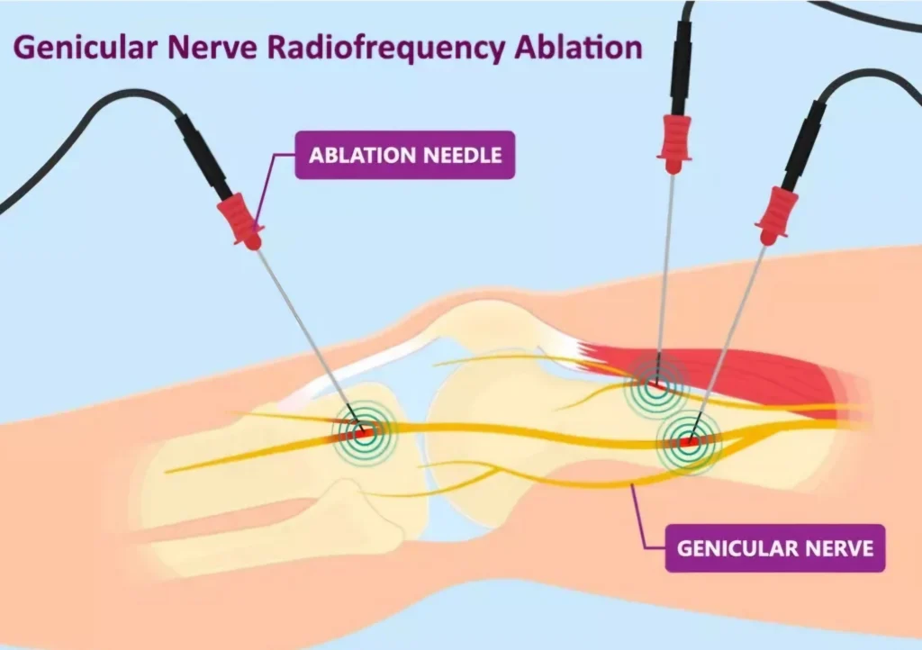
RFA is a minimally invasive percutaneous procedure, not open major spinal surgery. It involves no structural osteotomy, bone resection, or internal instrumentation/fusion. Utilizing specialized insulated radiofrequency needles, high-frequency electromagnetic waves generate targeted thermal coagulative necrosis to ablate the sensory medial branch nerves, effectively interrupting pain transmission to the brain without compromising the native biomechanical integrity of the spinal facet joint itself.
Clinical Advantages vs. Procedural Limitations
| Clinical Advantages of RFA (19.Radiofrequency Ablation RFA.pdf) | Limitations and Post-Procedural Considerations (19.Radiofrequency Ablation RFA.pdf) |
| • Puncture-sized percutaneous entry, minimal tissue trauma, avoiding major surgical exposure • Rapid functional recovery without extended inpatient hospitalization • Preserves native spinal architecture and biomechanics • Prevents or delays unnecessary spinal fusion surgeries • Safe to repeat upon eventual peripheral nerve regeneration | • Not an irreversible or permanent lifetime cure • Clinically significant analgesia typically persists for an average of 8–12 months • Transient post-procedural soreness or transient chemical neuritis may occur over the first 1–2 weeks • Peak therapeutic analgesic efficacy is typically realized between weeks 2 and 6 post-procedure |
Procedural Overview: The intervention is performed in an aseptic operating suite under real-time fluoroscopic guidance. The patient is placed prone and administered monitored local anesthesia with mild conscious sedation. The procedure duration is approximately 30–60 minutes, and the patient is discharged home on the same day.
Research References
- Chan CW, Peng P. Failed back surgery syndrome. Pain Medicine. 2011;12(4):577-606.
- Thomson S. Failed back surgery syndrome: definition, epidemiology and demographics. British Journal of Pain. 2013;7(1):56-59.
- Cohen SP, Raja SN. Pathogenesis, diagnosis, and treatment of lumbar zygapophysial (facet) joint pain. Anesthesiology. 2007;106(3):591-614.
- Schwarzer AC, Wang SC, Bogduk N, et al. Prevalence and clinical features of lumbar zygapophysial joint pain: a study in an Australian population with chronic low back pain. Annals of the Rheumatic Diseases. 1995;54:100-106.
- Manchikanti L, Manchukonda R, Pampati V, et al. Prevalence of facet joint pain in chronic spinal pain of cervical, thoracic, and lumbar regions. BMC Musculoskeletal Disorders. 2004;5:15.
- Cohen SP, Huang JH, Brummett C. Facet joint pain-advances in patient selection and treatment. Nature Reviews Rheumatology. 2013;9(2):101-116.
- Cohen SP, Hurley RW, Christo PJ, Winkley JV. Clinical predictors of success and failure for lumbar facet radiofrequency denervation. Clinical Journal of Pain. 2007.
- Cohen SP et al. Lumbar zygapophysial (facet) joint radiofrequency denervation success as a function of diagnostic block relief. Spine Journal. 2008.
- Cohen SP et al. Consensus practice guidelines on interventions for lumbar facet joint pain from a multispecialty, international working group. Regional Anesthesia and Pain Medicine. 2020;45(6):424-467.
- Provenzano DA et al. Assessment of real-world, prospective outcomes in patients treated with lumbar radiofrequency ablation for chronic pain (RAPID Study).
- Son JH, Kim SD, Kim SH, Lim DJ, Park JY. The efficacy of repeated radiofrequency medial branch neurotomy for lumbar facet syndrome. Journal of Korean Neurosurgical Society. 2010;48(3):240-243.